Arteriovenous Malformation (AVM)
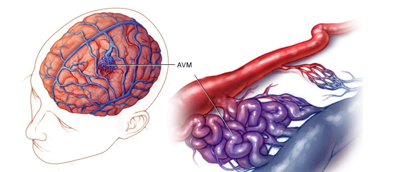
Arteriovenous malformation (AVM) is a tangle of abnormal and poorly formed blood vessels, primarily veins, linking arteries, and veins. AVMs usually develop in the brain or spinal cord but can also occur in other areas of the body. The abnormal vessels may cause clots to form that can lead to stroke or bleeding that’s difficult to control.

AVMs are most often diagnosed in adults between the ages of 30 and 60 who have symptoms such as seizures, headaches, and memory problems linked with bleeding episodes that happen without any trauma or injury.
No Capillaries
An arteriovenous malformation (AVM) is a tangle of abnormal and poorly formed blood vessel connections between veins and arteries such that there are no capillaries. AVMs are usually small in size but can be much larger. Most AVMs develop in the brain, spinal cord, or kidneys. Several different AVMs depend on where they’re located within the body.
AVM symptoms vary depending upon their location within the body and whether they cause bleeding into nearby tissues or cause pressure build-up inside the skull that leads to headaches or seizures if there’s a rupture in one part of an AVM leading to bleeding into another part of it; this is known as hemorrhage-induced stroke

Treatment options include endovascular coiling (or embolization), radiation therapy, or surgery, depending on how severe your condition is
AVMs pose a big risk.
The steal effect occurs when the increased blood flow through an AVM pulls oxygen away from your body’s normal tissues. This can impair the function of vital organs, including your brain and heart.
AVMs can also bleed into the brain or spinal cord, causing a stroke. Some AVMs grow larger over time and may eventually cause a heart attack or death.
Symptoms
AVMs are most commonly found in the brain but can also occur in the spinal cord. Symptoms may include:
- Headaches
- Dizziness or vertigo
- Weakness or paralysis
- Numbness, tingling, or loss of sensation in an area of your body
- Blurred vision or double vision (diplopia)
You should see a doctor immediately if you have any of these symptoms.
Diagnosis
To diagnose an AVM, doctors use a combination of techniques. They may conduct a CT scan, MRI, or both (depending on the type and location of the AVM). Angiography is also used to diagnose an AVM. This technique uses X-rays during injection of contrast agent into blood vessels to see if there are any abnormalities in blood vessels in your brain.
- Angio-CT: A procedure that combines conventional CT scans with contrast material to create pictures of arteries and veins within the brain
- Angio-MRI: A noninvasive test that combines magnetic resonance imaging (MRI) technology with enhanced images of blood vessels inside your head and neck

Treatment
Once diagnosed with an AVM, your doctor will advise you on the best course of treatment. Depending on the size and location of the AVM, surgical removal through a small incision may be advised. In this case, a surgeon can remove it either by clipping off its blood supply or completely removing it.
If surgery isn’t possible or doesn’t offer relief from symptoms, other options are available, including
- Endovascular embolization (using glue to plug off one of the arteries feeding it)
- Radiofrequency ablation (sending radio waves to destroy tissue)
- Stereotactic radiosurgery (using highly focused radiation)
Conclusion
AVMs are a rare but serious medical condition affecting millions worldwide. Thankfully, we have treatments to help manage the symptoms and prevent further complications. If you or someone in your family has been diagnosed with an AVM, please reach out to one of our experts today!


